
Microscope cameras are equipped with a one key white balance function primarily to address color bias in imaging and ensure accurate color reproduction of samples. Below are the specific reasons:
1. Instability of Light Source Color Temperature
– The color temperature of microscope light sources (e.g., halogen lamps, LEDs) fluctuates due to factors like usage time, voltage changes, or aging. For example, halogen lamps gradually shift toward red as they age, causing a warm (yellow/red) tint in images.
– One key white balance performs real-time calibration to counteract these shifts, neutralizing white areas (e.g., slide backgrounds or unstained sample regions) and maintaining color consistency.
2. Sample and Staining Specificity
-Different samples (e.g., H&E staining, fluorescent labeling) interact uniquely with light, potentially misleading the camera’s auto white balance. For instance, strong monochromatic light in fluorescence imaging can disrupt default RGB balancing.
-By manually selecting a reference point (e.g., an unstained area), users can correct color casts caused by the sample’s inherent properties.
3. Interference from Optical Path Components
-Filters, beam splitters, or other optical elements in the microscope’s light path may introduce color shifts (e.g., excitation filters in fluorescence microscopy enhance specific wavelengths).
-White balance compensates for these systematic biases, aligning imaging colors with the true sample appearance.
4. Limitations of Camera Sensors
-CMOS/CCD sensors exhibit varying sensitivity to different wavelengths (e.g., lower efficiency for blue light), which may result in a blue or green bias in raw images.
-White balance algorithms adjust RGB channel gains to match the sensor’s output to human perception or standardized references.
5. Rigor in Post-Analysis and Publication
-In research or medical diagnostics, color accuracy directly impacts interpretation (e.g., distinguishing pink vs. purple in histopathology slides may indicate different tissue states).
-One key white balance prevents misdiagnosis or data errors caused by color distortion, ensuring compliance with academic reproducibility standards.
How It Works
When the one key white balance is activated, the camera designates a user-selected area (e.g., a blank background) as the “white” reference. It then calculates the proportional differences among RGB channels and adjusts gains to neutralize the reference (R=G=B), thereby correcting all other colors.
Practical Applications
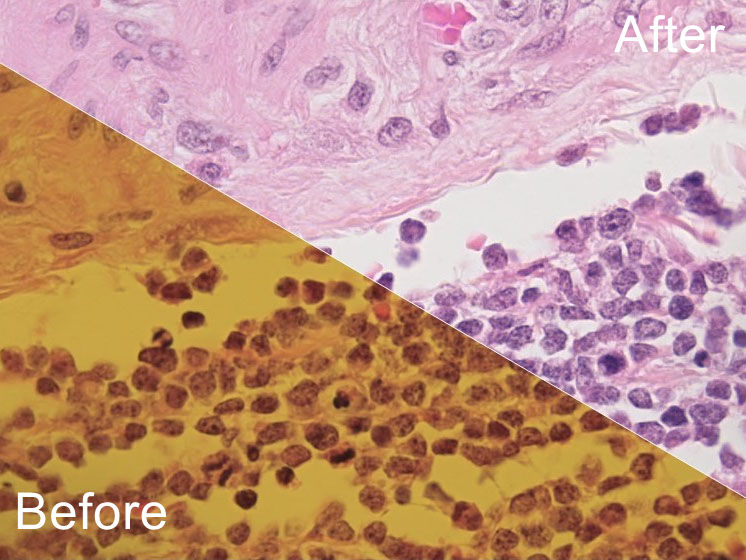
Pathology: H&E-stained slides require precise differentiation between nuclei (blue) and cytoplasm (pink); color shifts could lead to diagnostic errors.
Fluorescence Imaging: Corrects crosstalk between fluorescent channels, preventing signal bleed-through.
Time-Lapse Imaging: Compensates for color temperature drift caused by prolonged light source operation.
Conclusion
The one key white balance in microscope cameras is a critical tool for color management. By addressing biases introduced by light sources, samples, optical systems, and sensors, it ensures scientific accuracy and visual reliability. For applications demanding precise color fidelity (e.g., medicine, materials science), this feature is indispensable.