
Introduction
The modern microscope is no longer confined to the eyepiece. The integration of a high-quality microscope camera has transformed optical instruments into powerful digital imaging stations, unlocking new levels of analysis, documentation, and sharing. From cutting-edge research labs to industrial quality control and educational classrooms, these cameras are indispensable tools. This guide explores the core applications and key benefits of microscope cameras, explaining why they are a critical upgrade for any microscopy setup.
What is a Microscope Camera?
A microscope camera, or digital microscopy camera, is a specialized imaging device designed to attach to a microscope’s photo port. It captures high-resolution still images and video of specimens, converting the optical view into digital data. Key components include a sensitive image sensor (CMOS or CCD), suitable optics, and software for control and processing.
Core Applications of Microscope Cameras
Microscope cameras serve vital functions across diverse sectors:
1. Research & Life Sciences
Documentation & Analysis: Precisely capture cell structures, tissue samples, or fluorescently labeled specimens for quantitative analysis (e.g., measuring cell count, area, fluorescence intensity).
Time-Lapse Studies: Record dynamic processes like cell division, growth, or movement over hours or days.
Sharing & Collaboration: Easily share images with colleagues worldwide for peer review or collaborative diagnosis.
2. Medical & Clinical Diagnostics
Telepathology: Transmit real-time images of blood smears, tissue biopsies, or cytology samples for remote consultation and diagnosis.
Digital Record-Keeping: Create permanent, searchable patient records by archiving digital slides and samples.
Education & Training: Capture classic case studies for teaching medical students and trainees.
3. Industrial Quality Control & Failure Analysis
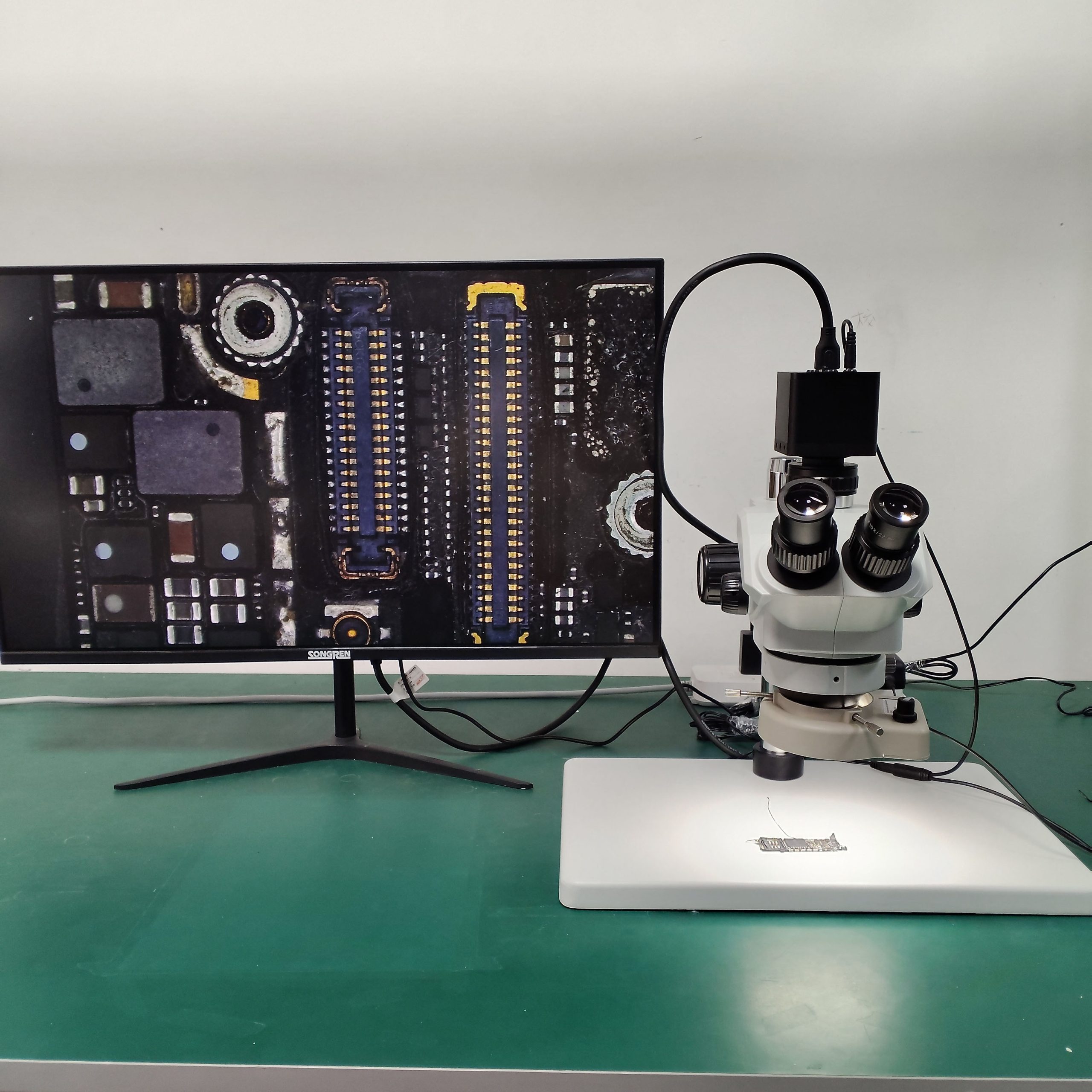
Inspection & Measurement: Document defects, measure coating thicknesses, analyze material fractures, or inspect solder joints on circuit boards with precise calibration tools.
Reporting: Generate visual evidence for audit trails, client reports, and compliance documentation (e.g., in semiconductor, aerospace, metallurgy).
Team Review: Share inspection results instantly across production, engineering, and quality teams for faster decision-making.
4. Education & Academia
Enhancing Learning: Project live microscope views to an entire classroom, ensuring all students see the same detailed image.
Student Projects: Allow students to capture, annotate, and submit their own digital images for assignments and assessments.
Creating Learning Materials: Build libraries of reference images for coursework and online learning modules.
5. Forensic Science
Evidence Documentation: Create incontrovertible digital records of trace evidence like fibers, gunshot residue, or document authenticity.
Analysis: Perform detailed comparisons of materials or tool marks.
Courtroom Presentation: Present clear, high-resolution visual evidence to juries and officials.
Key Benefits & Why You Need a Microscope Camera
Integrating a camera delivers transformative advantages:
Enhanced Visualization & Analysis: Specialized software enables measurements, annotations, and image enhancement (adjusting contrast, sharpness) beyond the capabilities of the human eye alone.
Superior Documentation: Create a permanent, organized digital archive. No more relying on hand-drawn sketches or fading physical photos.
Improved Efficiency & Workflow: Streamline reporting, share findings in seconds, and facilitate remote work and consultations.
Quantitative Data Acquisition: Extract precise, reproducible numerical data from your samples, essential for statistical analysis and publication-grade research.
Accessibility & Collaboration: Make microscopy accessible to groups and break down geographical barriers for expert input.
Choosing the Right Microscope Camera
Consider these factors:
Sensor Resolution (Megapixels): Higher resolution captures finer detail but requires more processing power.
Sensor Sensitivity & Type: A sensitive CMOS or CCD sensor is crucial for low-light applications like fluorescence microscopy.
Frame Rate: For smooth video of moving specimens, a higher frame rate (e.g., 60 fps or more) is necessary.
Software Capabilities: Ensure the included software supports your needs (measurement, stitching, extended depth of focus, etc.).
Compatibility: Verify the camera mount (C-mount standard) and software are compatible with your microscope model and computer system.
Conclusion
A microscope camera is more than an accessory; it’s a gateway to advanced digital microscopy. It elevates your work from simple observation to comprehensive analysis, documentation, and communication. Whether for rigorous scientific research, precise industrial inspection, or dynamic educational instruction, investing in the right microscope camera enhances accuracy, productivity, and the overall impact of your work.