
Introduction
What is a Microscope Camera?
A microscope camera, often termed a digital imaging system, is a device mounted on a microscope to capture, display, and save high-resolution still and video images of specimens. These cameras connect to a computer monitor, allowing multiple professionals to view the same sample simultaneously and perform detailed analysis using specialized software.
Key Applications of Microscope Cameras in Medical Diagnosis
The use of digital imaging is pervasive across medical fields that rely on microscopic analysis.
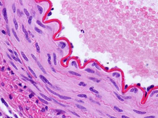
1. Digital Pathology and Histopathology
This is perhaps the most significant application. Microscope cameras enable the creation of whole-slide images (WSI)—high-resolution digital scans of entire glass slides. Pathologists can then:
-Diagnose diseases like cancer from a digital screen instead of a traditional microscope.
-Annotate specific areas of interest directly on the image.
-Store vast digital archives for easy retrieval and review.
2. Hematology
In blood analysis, cameras are essential for:
-Capturing images of blood smears for identifying abnormal cells, parasites (e.g., malaria), and disorders like leukemia.
-Documenting rare findings for training purposes and second opinions.
-Sharing critical results with oncologists or other specialists quickly.
3. Microbiology and Cytology
Microbiologists use cameras to:
-Identify bacteria, fungi, and viruses based on morphological characteristics.
-Record time-lapse videos of cell culture growth and behavior.
-Document Pap smears and other cytological samples for cancer screening.
4. Intraoperative and Surgical Consultation
During complex surgeries, such as tumor removals, a frozen section analysis is often performed. A microscope camera allows:
-The pathologist to stream live images to the surgical team in the operating room.
-Surgeons to see the microscopic findings in real-time, enabling immediate decision-making about the surgical margins.
5. Telepathology and Remote Consultation
Microscope cameras break down geographical barriers. Specialists in one part of the world can:
-Easily share images with colleagues globally for a second opinion.
-Provide diagnostic services to remote or underserved clinics lacking expert staff.
-Collaborate on complex cases in real-time.
The Importance and Benefits: Why They Are indispensable
The adoption of microscope cameras is not just a technological upgrade; it’s a fundamental shift in diagnostic medicine.
-Enhanced Diagnostic Accuracy: Digital images allow for magnification, contrast adjustment, and precise measurement on a large screen, reducing eye strain and potential human error associated with prolonged microscope use.
-Efficient Documentation and Archiving: Images and videos become a permanent part of the patient’s electronic health record (EHR). This creates an invaluable resource for tracking disease progression, comparing past and present samples, and auditing.
-Improved Collaboration and Training: Multiple people can view the same high-quality image simultaneously. This is crucial for teaching medical students, training new technicians, and discussing cases in multidisciplinary team meetings.
-Streamlined Workflows and Increased Productivity: Digital pathology slides can be managed, sorted, and analyzed faster than physical slides. Automated software can even assist with pre-screening and counting cells.
-Objective Data Analysis: Image analysis software can provide quantitative data (e.g., cell count, staining intensity, tumor area) that is more objective and reproducible than subjective visual estimates.
Microscope cameras have evolved from a simple documentation tool to an integral component of the diagnostic pipeline. Their ability to enhance accuracy, improve collaboration, and create digital workflows makes them indispensable in modern medicine. As technology advances, particularly with the integration of Artificial Intelligence (AI) for automated image analysis, the role of the microscope camera will only grow more critical, paving the way for faster, more precise, and more accessible patient diagnosis worldwide.